Suspect ADH1:
Uncover the mystery

What is ADH1?

Autosomal dominant hypocalcemia type 1 (ADH1) is a rare genetic condition but a common cause of nonsurgical hypoparathyroidism.1

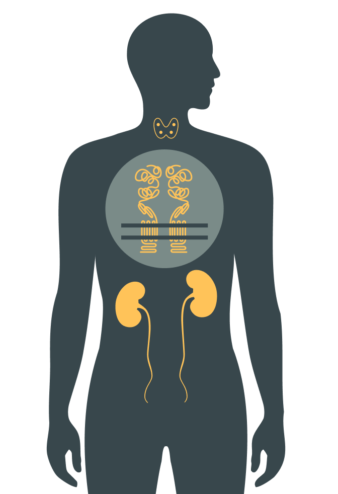
It is an inherited endocrine disorder characterized by2:

  • Decreased parathyroid hormone (PTH) secretion
  • Low serum calcium concentration
  • Increased urinary calcium excretion
Circle icon with forest green background featuring 4 people icons in gray and 1 in yellow with text: ADH1 affects up to 1 in 25,000 people
Info Icon

A child has a 50% chance of inheriting the variant associated with ADH1 if one parent is affected.2

Icon in forest green of human body featuring gold thyroid and kidneys, and grey circle with gold process in chest

ADH1 is a distinct form of hypoparathyroidism

Hypoparathyroidism can result from surgical or nonsurgical etiologies, which include genetic disorders.1 ADH1 is a genetic form of nonsurgical hypoparathyroidism caused by gain‑of‑function variants in the calcium-sensing receptor gene (CASR), of which over 100 are known.2

Variants in the calcium-sensing receptor (CaSR) are responsible for1,2:

  • Reduced PTH secretion
  • Reduced renal calcium reabsorption

The mechanism of disease in ADH1 is distinct from that of other forms of hypoparathyroidism because of the critical role of the CaSR in both the parathyroid glands and kidneys.2,3

ADH1 symptoms and impact on patients2

A common sign of ADH1 is low serum calcium, which results in symptoms including tetany, muscle cramps, and spasms. In more severe cases, seizures, laryngospasms, and cardiac arrhythmias are experienced. Conventional treatment (calcium and active vitamin D supplementation) can lead to kidney calcification, kidney stones, and even kidney failure.  

Physical symptoms of ADH1

  • Muscle cramps and spasms (tetany)
  • Seizures
  • Laryngospasms
  • Heart rhythm disorders

Long-term complications*

  • Kidney calcification (nephrocalcinosis)
  • Kidney stones (nephrolithiasis)
  • Kidney failure

*Result of prolonged conventional treatment with calcium supplements and active vitamin D analogues.

Icon of dark green human body showing bones, heart, kidneys, and urethra.6 symbols in light green surround the body depicting brain, sleep, emotions, and bone
Collection of icons include blue cloud with raindrop, human head with puzzle piece and heart rhythm and triangle with exclamation point

The hidden burden of ADH12

People living with hypoparathyroidism, including ADH1, may experience these quality-of-life challenges:

  • Fatigue
  • Depression
  • Cognitive impairment
  • Anxiety
Info Icon

Some people with ADH1 never learn of the underlying genetic cause of their condition and may never receive optimal disease management.

Impacts of diagnostic delay2

While symptoms may start in infancy, many people with ADH1 may not receive an accurate diagnosis until they are adults.

Calendar graphic with 20+years and triangle with exclamation point.

Twenty-plus-year gap between median age of diagnosis for a hypocalcemia-related disorder (4 years) and genetic confirmation of ADH1 (25 years)

Gold circle with 25% black outline and head and shoulder person icon with 75%

Treatment-emergent complications occurred in 75% of patients evaluated

Patients may receive an incomplete diagnosis, including:

  • Idiopathic hypoparathyroidism
  • Hypocalcemia

Patients may receive a misdiagnosis based on symptoms:

  • Seizure disorder
  • Respiratory-related disorder
  • Cardiovascular-related disorder
DNA Icon

Do you suspect ADH1?

Genetic testing is recommended for patients with idiopathic hypoparathyroidism.

What causes ADH1?

Video overview: Understanding ADH1

Gain‑of‑function variants in the CASR gene are the root cause of ADH12

These CASR variants cause the CaSR to be overly sensitive to calcium, thereby causing it to detect low blood calcium concentration as "normal." This leads to decreased PTH secretion from the parathyroid glands and decreased calcium reabsorption in the kidney—the result is:

  • Low blood calcium concentration (hypocalcemia) and high (hypercalciuria) or inappropriately normal urine calcium concentration
black header 3 circles featuring blood system, parathyroid and kidneys.
gold header with 3 circles featuring blood system in gold, parathyroid with red dots, and kidneys in gold
Icon of 3 symbols: gold kidneys, 3 blue drops, and a black triangle outline with exclamation point

ADH1 and the risk of kidney damage

Due to the critical role of CaSR on the functions of the parathyroid gland and kidneys, patients with ADH1 often present with both hypocalcemia and hypercalciuria and may experience hypercalciuria-associated complications2,4:

  • Nephrocalcinosis
  • Nephrolithiasis
  • Renal impairment

Current management of ADH1

For people living with ADH1, the main goal with conventional therapy is to keep blood calcium concentrations close to, or slightly below, the lower limit of normal to avoid2,3,5:

  • Symptomatic hypocalcemia
  • Hypercalciuria
  • Associated renal complications

For patients with chronic hypoparathyroidism, current guidelines suggest these tests at baseline and every 3–6 months5-7:

Blood tests

Serum calcium
(albumin-adjusted or ionized)

Magnesium

Phosphorus

25-hydroxyvitamin D

Serum creatinine (for eGFR)

Urine tests

24-hour urine creatinine

24-hour urine calcium

eGFR=estimated glomerular filtration rate.

Info Icon

Patients with ADH1 should be monitored regularly to assess their blood biochemistry and the risk of hypercalciuria and potential kidney damage.2,6

Currently, there are no medications specifically indicated to treat ADH1. There remains a substantial unmet need for therapies that address the underlying molecular mechanism of ADH1.2,5

Inform ADH1 management with a confirmatory genetic test

References: 1. Mannstadt M, Cianferotti L, Gafni RI, et al. Hypoparathyroidism: genetics and diagnosis. J Bone Miner Res. 2022;37(12):2615-2629. 2. Roszko KL, Stapleton Smith LM, Sridhar AV, et al. Autosomal dominant hypocalcemia type 1: a systematic review. J Bone Miner Res. 2022;37(10):1926-1935. 3. Roszko KL, Bi RD, Mannstadt M. Autosomal dominant hypocalcemia (hypoparathyroidism) types 1 and 2. Front Physiol. 2016;7:458. 4. Pearce SH, Williamson C, Kifor O, et al. A familial syndrome of hypocalcemia with hypercalciuria due to mutations in the calcium-sensing receptor. N Engl J Med. 1996;335(15):1115-1122. 5. Khan AA, Ali DS, Bilezikian JP, et al. Best practice recommendations for the diagnosis and management of hypoparathyroidism. Metabolism. 2025;171:156335. 6. Bollerslev J, Rejnmark L, Marcocci C, et al. European Society of Endocrinology clinical guideline: treatment of chronic hypoparathyroidism in adults. Eur J Endocrinol. 2015;173(2):G1-20. 7. Bollerslev J, Buch O, Cardoso LM, et al. Revised European Society of Endocrinology clinical practice guideline: treatment of chronic hypoparathyroidism in adults. Eur J Endocrinol. 2025;193(5):G49-G78.